
Leeds Centre for Disease Models image competition winners
🎉 Huge congratulations to Erica Harris, Basudha Basu, Magdalena Karwatka and Marine Krzisch for claiming first, second, and third place in the LCDM Image Competition, proudly sponsored by Proteintech!
🏆 Erica dazzled the judges and took home a £50 Amazon voucher, while all three winners received the coveted Proteintech alpaca — a prize almost as legendary as their stunning submissions.
📸 The images they submitted were nothing short of extraordinary — a brilliant showcase of creativity, precision, and scientific beauty. Well done to all!

iPSC-derived Astrocytes after 44 days in culture, staining for cytoplasmic glial fibrillary acidic protein GFAP (green) and mature astrocyte/oligodendrocyte marker S100B (red)
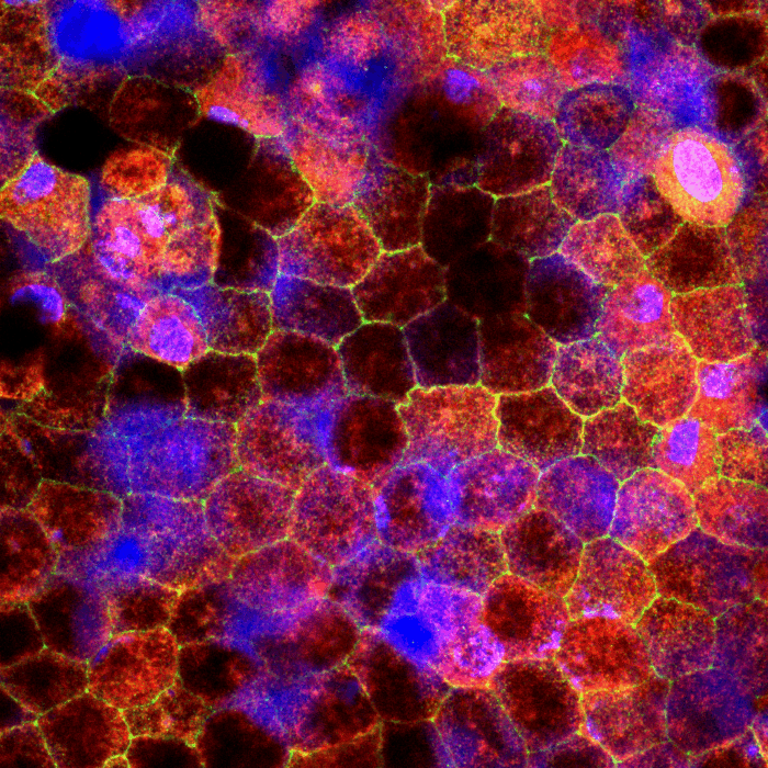
Human iPS cell line was differentiated into retinal pigment epithelial (RPE) cells using a series of biochemical factors. RPE cell layers have a characteristic cobblestone appearance and are marked by the five "P"s - polygonal, polarized, postmitotic, phagocytic and pigmented. In this image, ZO-1 (in green) marks the tight junctions in the cell boundaries emphasizing the cobblestone appearance. Some heavily melanin-pigmented cells are also visible. RPE-65 (stained in red) is a major player of the visual cycle which regenerates visual pigments in rod and cone photoreceptors. Together these antibodies mark mature RPE cells. DAPI, marking the nucleus, is in blue.

Kidney organoid differentiated from induced pluripotent stem cells (iPSCs) carrying a patient-specific mutation causing Joubert syndrome. The organoid has been stained for kidney markers: CDH1 (blue) for distal tubules and collecting ducts, LTL (green) for proximal tubules, and NPHS1 (red) for podocytes. Visible cysts are present along the edges of the organoid.
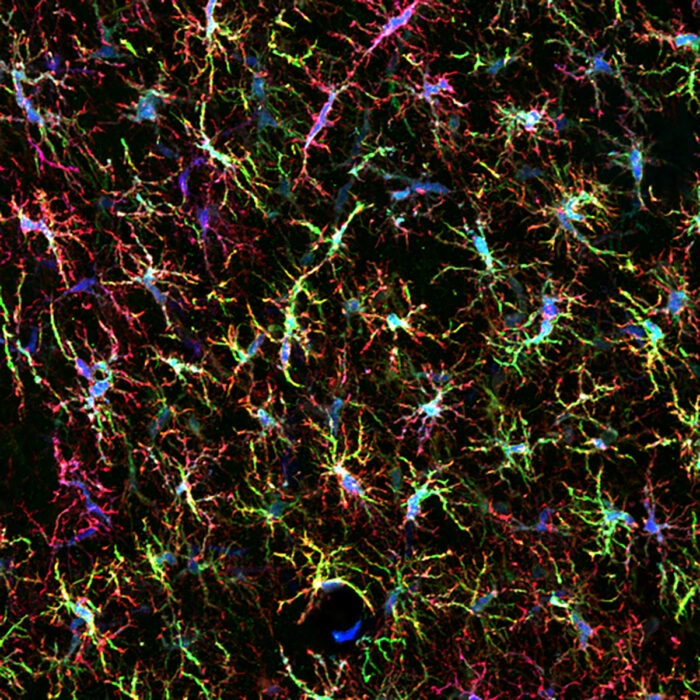
We transplanted myeloid precursors derived from familial Parkinson’s disease patients and from patients where the mutation was corrected into the mouse brain. These precursors migrated throughout the brain and differentiated into microglia. This picture shows Parkinson patient-derived GFP-labeled microglia (green) in the mouse brain at 1 month post-injection. They were labeled with the microglial markers P2RY12 (blue) and Iba1 (red).